Subject: Biology
Grade Level: 9th
grade
Lesson Duration: 75 minutes
Benchmark:
5c Cells: Before
a cell divides, the instructions are duplicated so that each of
the two new cells gets all the necessary information for carrying
on.
State Benchmark: 12.A.4b
Describe the structures and organization of cells and tissue that underlie basic life functions including…reproduction. [Sample benchmark indicator: Explain asexual reproduction (mitosis)]
Objectives:
1. The students will
be able to identify specific stages of mitosis from illustrations
and from animal cells on slides under a microscope.
2. The students will
be able to draw and label a cell in each stage of mitosis
(prophase, metaphase, anaphase, and telophase).
3. The students will
be able to explain how both of the new cells produced in mitosis
contain identical genetic information.
4. Students should be
able to predict what would be happen if there were an error in
mitosis.
Students
Background:
The students should have
a solid understanding of the structure of a cell. Students
need to know what chromosomes are and understand that they carry
the genetic information of an individual. They also need to
know that every cell in an organism must have the same
chromosomal DNA. I would precede this lesson with a
discussion on why cells need to divide. The students must
also have a working knowledge of how to properly use a
microscope. Students will also need to have pre-assigned
lab partners and lab jobs.
Materials Needed:
Activity
Overview:
This lesson is designed
to teach students the steps of mitosis and how the genetic
material changes and moves to create two new identical cells.
There are three different activities in this lesson.
First, the class will do
the sock activity. This activity will help the students see
how the chromosomes of the cell move throughout mitosis and
finally end up with two genetically identical cells. Chromosomes
(visibly different pairs of socks) will be in the nucleus (a
large circle on the floor) of a cell. The class will assume
that the cell has already replicated its chromosomes. Each
pair of socks will be a pair of chromatids. (Note: Do
not fold the socks as you would if you had just done the laundry.
Simply place them next to each other so that the students can see
that they are paired.)
Second, the class will
watch a computer animation of mitosis. I will hook the
computer up to a TV or projector so that all the students can
watch at the same time. I will go to the website: http://www.sci.sdsu.edu/multimedia/mitosis/. This particular animation can be paused,
watched frame by frame, or sped up. This will allow me to
point out things in the middle of the animation and allow
students to ask questions. At the same website, there is a
video of an actual cell dividing. I will show this several
times as well.
Last, the students will pair up and observe prepared slides of the stages of mitosis under a light microscope. They will identify and sketch out what they see. A worksheet with examples of each of the stages of mitosis will be provided. The students will take these sheets home and briefly explain what is happening in each stage.
Introduction:
(10 minutes)
Teacher (T): Last
class we were discussing cells and why they need to divide.
Who can tell me why our cells divide? (I will allow wait
time after every question.)
Student (S): Well,
we are made of trillions of cells and we only start out from one.
The cells must divide for us to grow.
T: That’s
good. Are there any other reasons why cells might divide?
S: Sometimes cells
die and we need to make new ones.
T: That could be
another reason. There is one more major reason that a cell
might divide, can anyone tell me what it is?
S: Cells have a
volume to surface area ratio that they must keep low. If
the cell gets too big in volume, then it has too little surface
area for it to let enough things in and out of the cell and it
would not work very well.
T: Very good!
So we know that cells need to divide. Let’s imagine
that we have a cell that has reached its volume to surface area
ratio and it needs to divide to remain properly functional.
This cell needs to divide up evenly and both new cells need all
of the same things that the large cell has. This means that
somehow, this cell needs to make sure that each new cell has the
same genetic information. If they do not have the exact
same genetic information, the cell will not function properly.
How do cells do this? That’s what we are going to
discover today!
Body of Lesson:
Sock Activity
(20 minutes)
T: If everyone
could come over to this side of the room we will begin our first
activity.
» Have
the 10 (or however many) visibly different pairs of socks
scattered on the floor within a large circle of tape (the cell
wall). Have matching pairs next to each other, but not
folded together. I will also have a copy of the
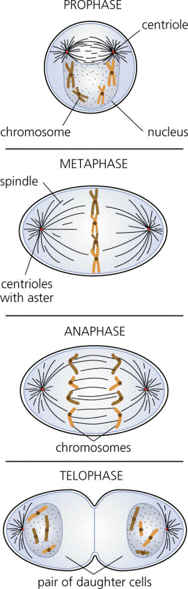
stages of mitosis (appendix
1) on the overhead. I will
cover the stages with a sheet of paper and uncover them as we go
along.
T: This is our cell
that needs to divide. The different socks are the
cell’s different chromosomes. Can someone remind the
class what chromosomes are?
S: Chromosomes are
condensed pieces of DNA in a cell.
T: That’s
right. The chromosomes in this particular cell have already
replicated or copied themselves. As you can see, each
chromosome is made of two identical pieces. These are the
identical copies of the cells DNA. There are many different
chromosomes and they all are made of 2 identical copies of DNA.
If you look at the overhead, you can see that this stage of cell
division is called prophase. Now before we go on, can
someone tell us what each of these socks represent?
S: Each pair of
matching socks is a chromosome. Each chromosome is made of
two identical copies of the same DNA. That is what each
individual sock is.
T: Exactly. So
now we can move on to the next step. In this step, all of
the chromosomes line up down the middle of the cell like this.
(I will move the socks to line up down the middle of the circle.)
This stage is called metaphase. (I will uncover metaphase
on the overhead.) This is getting the chromosomes ready to
split up and move to either side of the cell. So now, what
happens in metaphase?
S: The chromosomes
line up down the center of the cell.
T: Yes. Now
we can see what happens to the chromosomes. An apparatus in
the cell is formed called the spindle fibers. The spindle
fibers are just like pieces of string from either side of the
cell that reach out to each chromosome and pull the identical
pieces in opposite directions. The spindle fibers attach to
the centromere, or the center of each chromosome. Then,
they begin to pull half of the chromosome to each side of the
cell. (I will begin to separate the pair of socks by moving
one sock of each pair in either direction.) This stage is
called anaphase. (I will uncover anaphase on the overhead.)
Okay, there were some key terms and ideas in this stage. First,
can someone explain what the spindle fibers do?
S: The spindle
fibers come from two opposite ends of the cell and reach out and
pull the chromosomes apart.
T: That’s
good. Does anyone remember where the spindle fibers attach
to the chromosomes?
S: The center of
the chromosome. That’s called the centromere.
T: Very good.
You can see the centromeres more clearly on the overhead. There
are the little circles in the center of the chromosomes. (I
will point them out). Now the chromosomes are separating
out equally to opposite sides of the cell. The chromosomes
finish moving and then the cell actually needs to divide into two
separate cells. (I will finish moving the chromosomes apart
and then put a new piece of tape down the middle separating it
into two new cells.) This stage is called telophase. (I
will uncover telophase on the overhead.) All of these
stages together are what happens during cell division. All
of these steps together is called mitosis. So we have gone
through four different steps to mitosis. Can someone start
us at the beginning and tell us what happens? Feel free to
show us with the socks.
S: Well, first it
starts in prophase where the DNA is already replicated and is
condensed into chromosomes.
T: And what are the
chromosomes composed of?
S: Two identical
copies of DNA. Each sock in the chromosome represents a
copy.
T: Great. So
we have replicated DNA in the form of chromosomes. Can
someone else tell us what happens next?
S: The chromosomes
line up down the middle in the next step called metaphase.
T: Exactly. And
then what happens?
S: The chromosomes
separate into the two separate copies and move to opposite sides
of the cell.
T: Great. What
are the things that pull the chromosomes apart called?
S: Spindle fibers.
T: And the spindle
fibers attach where on the chromosome?
S: The centromere.
T: Exactly. And
this all happens in which step?
S: Anaphase.
T: What happens
after anaphase?
S: Telophase.
T: Yes, that is
right, and what happens in telophase?
S: The chromosomes
finish dividing and the cell splits into two separate cells.
T: That’s
right. And what happens after that?
S: That’s the
end.
T: Well, it’s
the end of this cell division, but the cell will work and grow
and then it will need to divide. After awhile each of these
new cells will go through mitosis too! Now that we have
looked at mitosis using socks, we are going to see a short
animation and video of mitosis. Please take your seats.
Computer Animation and
Video: (10 minutes)
» I would set up the computer and TV or projector
ahead of time. I would also test it to make sure that
everything is working well. The website address is: http://www.sci.sdsu.edu/multimedia/mitosis/
T: Now I have an
animation that we will watch. First we will watch the whole
thing. It shows us the stages of mitosis. Now before
we start, let’s make sure that we all understand what we are
looking at. Can someone tell me what the red and blue
x’s in the middle are?
S: They are the
chromosomes.
T: Right. And
like the pairs of socks, they are already replicated or copied.
You can see two halves on each chromosome. Those are the
identical copies of DNA. Now let’s watch the whole
thing.
» I will show the animation in its entirety. I
would show it at about 2 frames per second. At this speed,
I can tell the students which stage the cell is in.
T: I hope everyone
was able to follow along. Now we can go through it frame by
frame and talk about what it happening.
» I will start going through the animation frame by
frame.
T: When the
chromosomes are moving toward the center, which step is this?
(I would have the animation stopped on a frame showing
metaphase.)
S: Metaphase.
T: (Continue with
the animation frame by frame.) Now what is pulling the
chromosomes apart here in anaphase?
S: The spindle
fibers.
T: And where do the
spindle fibers attach?
S: The centromeres.
T: Exactly. You
can see the centromeres here. And you can see them
splitting up as the chromosomes separate. (Continue with
animation.) And now at then end of telophase, what do we
have?
S: Two genetically
identical new cells.
T: Good job class.
You seem to be understanding mitosis. The other part of
this animation is a video of a real cell undergoing mitosis.
Let’s watch that. (I will show the video of mitosis.)
I know that went by really fast, but that’s what it looks
like in a real cell. Let’s watch it again. (I
would show the video 2 or 3 more times depending on student
interest.) Great, now we can look at some slides of real
cells going through mitosis.
Microscope Activity:
(30 minutes)
T: You need to sit
next to your lab partner and have the materials person go get you
two a microscope. The other partner needs to get out two
sheets of paper. I will hand out the slides.
» Allow
time for students to get their microscopes. Hand out the
pre-made slides of the stages of mitosis.
T: Now that you all
have microscopes and paper, you all should be able to observe the
different stages of mitosis on your slide. Go ahead and
look at your slide and see what is on it. (I will pause for
the students to focus on their slides.) There are 4
separate cells on the slide. Each cell is frozen in a
specific stage of mitosis. I want you to sketch out each
stage on a sheet of paper. The sketch doesn’t need to
be very detailed, I simply what you to identify where the
chromosomes are in each of the stages and what they look like.
I will come around to each group to see if you are finding the
stages and to see your drawings. What is it that you need
in your sketches?
S: We need to draw
the cell and where the chromosomes are in each stage.
T: Right. You
can just draw four circles and then fill them in with the
positions of the chromosomes. Go ahead and get started.
I will be coming around.
» I will walk
around the classroom making sure that everyone is on task.
I will check the microscope image to make sure that they are
seeing what they need to be seeing. I will also look at
their sketches and help them if they are not doing it right.
Closure: (5
minutes)
T: For one last
time, let’s walk through the stages of mitosis using
everything we have learned today. Can someone tell me what
happens first in prophase?
S: In prophase, the
DNA has already copied itself and is condensed into chromosomes.
The chromosomes are made of identical copies of DNA.
T: Good, and then
what happens next?
S: The chromosomes
line up down the middle in metaphase.
T: That’s
right. What happens next?
S: Anaphase is
next. The spindle fibers attach to the centromeres of the
chromosomes and begin to pull the chromosomes apart. One
copy of each chromosome goes to each opposite side of the cell.
T: Great! Who
can tell me what happens after that?
S: The chromosomes
reach opposite ends of the cell and then the cell splits apart
and makes two new cells. This is called telophase.
T: You all really
seem to understand this. What is so special about the two
new cells that have been made?
S: They have
identical genetic material.
T: Wonderful!
It seems like you all have a fairly good grasp on mitosis now.
For your homework, I want you all to take the sketches that you
have drawn from the slides, and write a short explanation of what
is happening with the chromosomes in each step. Also
be sure to write the name of the stage of each sketch if you
haven’t already. That will be due at the beginning of
the next class. Have a great day!
Assessment:
1.
What is the purpose of mitosis? What are the end products?
(be specific)
2. Here are the four
stages of mitosis (I would have unlabeled diagrams of prophase,
metaphase, anaphase and telophase out of order.) Please
name them and put them in order (1-4).
3. Imagine that a cell
is going through mitosis. During anaphase, one of the
chromosomes does not separate properly. The entire
chromosomes is pulled to one side of the cell. This new
cell that forms from this side has the entire chromosome (both
identical copies of DNA). What might happen to the other
cell that is produced in this particular mitotic division?